Abstract
 |
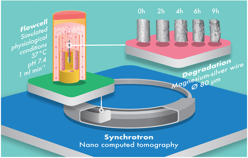
Functional materials feature hierarchical microstructures that define their unique set of properties. The prediction and tailoring of these require a multiscale knowledge of the mechanistic interaction of microstructure and property. An important material in this respect is biodegradable magnesium alloys used for implant applications. To correlate the relationship between the microstructure and the nonlinear degradation process, high-resolution in situ three-dimensional (3D) imaging experiments must be performed. For this purpose, a novel experimental flow cell is presented which allows for the in situ 3D-nano imaging of the biodegradation process of materials with nominal resolutions below 100 nm using nanofocused hard X-ray radiation from a synchrotron source. The flow cell setup can operate under adjustable physiological and hydrodynamic conditions. As a model material, the biodegradation of thin Mg-4Ag wires in simulated body fluid under physiological conditions and a flow rate of 1 mL/min is studied. The use of two full-field nanotomographic imaging techniques, namely transmission X-ray microscopy and near-field holotomography, is compared, revealing holotomography as the superior imaging technique for this purpose. Additionally, the importance of maintaining physiological conditions is highlighted by the preliminary results. Supporting measurements using electron microscopy to investigate the chemical composition of the samples after degradation are performed. |
Jan Reimers et al., Development of a Bioreactor-Coupled Flow-Cell Setup for 3D In Situ Nanotomography of Mg Alloy Biodegradation, ACS Appl. Mater. Interfaces