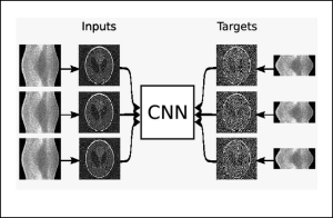
High-resolution X-ray nanotomography is a quantitative tool for investigating specimens from a wide range of research areas. However, the quality of the reconstructed tomogram is often obscured by noise and therefore not suitable for automatic segmentation. Filtering methods are often required for a detailed quantitative analysis. However, most filters induce blurring in the reconstructed tomograms. Machine learning (ML) techniques offer a powerful alternative to conventional filtering methods. Self-supervised denoising ML technique can be used in a very efficient way for eliminating noise from nanotomography data, outperforming conventional filters, such as a median filter and a nonlocal means filter without blurring relevant structural features.
Below find a list of selected denoising implementions readily available on the Maxwell cluster, and MDLMA publications touching this area.
Also including are more general purpose utilities for image manipulation and visualization
Applications
- denoiseg: Joint Denoising and Segmentation
- divnoising: Diversity Denoising with Fully Convolutional Variational Autoencoders
- histolab: WSI processing in deep learning pipelines
- napari: fast, interactive, multi-dimensional image viewer for Python
- noise2inverse: Self-supervised deep convolutional denoising for linear inverse problems in imaging
- pyg: PyG is a library built upon PyTorch to easily write and train Graph Neural Networks for a wide range of applications related to structured data.
- tomopy: Image reconstruction algorithms for tomography
- voxelmorph: general purpose library for learning-based tools for alignment/registration and modelling with deformations
Publications
- Reconstruction, processing and analysis of tomography data at the Hereon beamlines P05/P07 at PETRA III (Conference Presentation): Julian P. Moosmann et al., SPIE Optical Engineering + Applications, doi: 10.1117/12.2637973
- Machine learning denoising of high-resolution X-ray nanotomography data: Silja Flenner et al., J Synchrotron Radiat, doi: 10.1107/S1600577521011139
- Artifacts suppression in biomedical images using a guided filter: Inna Bukreeva et al., Thirteenth International Conference on Machine Vision, doi: 10.1117/12.2587571
